Mouth
• Formed by the cheeks, hard and soft palates, lips, and
tongue
• The vestibule: Space bounded externally by the cheeks and
lips and internally by the teeth and gums
• The tongue:
– Together with its associated muscles, forms the floor of
the oral cavity
– Composed of skeletal muscle covered with mucous membrane

Salivary Glands
• Secretes saliva
• Outside the mouth and pour their contents into ducts that empty
into the oral cavity
• 3 pairs of major salivary glands:
– Parotid
– Submandibular
– Sublingual glands
• Saliva lubricates food and starts the chemical digestion
of carbohydrates

Small
Salivary Glands
• The mucous membrane of the mouth and tongue contains many
small salivary glands
• Open directly or indirectly via short ducts to the oral
cavity
• Make a small contribution to saliva
• These glands
include:
– Labial glands – in lips
– Buccal glands– in cheeks
– Palatal glands – in palate
– Lingual glands – in the tongue
Composition
of Saliva
• Chemically, saliva is 99.5% water and 0.5% solutes
• Solutes:
– Ions including sodium, potassium, chloride, bicarbonate
& phosphate
• Dissolved gases and various organic substances
– Urea
– Uric acid
– Mucus
– Immunoglobulin A
• Bacteriolytic enzyme – lysozyme
• Salivary amylase – digestive enzyme that acts on starch
Functions
of Saliva
• Water in saliva:
Provides a medium for dissolving food
• Chloride ions:
Activate salivary amylase
• Bicarbonate and
phosphate ions: Buffer acidic foods that enter the mouth
• Mucus:
Lubricates food
• Immunoglobulin A
(IgA) – Prevents attachment of microbes
• Lysozyme –
Kills bacteria
Salivation
• Control: By the autonomic nervous system
• Amounts of saliva –
1000–1500 mL
• Parasympathetic
stimulation
– Promotes continuous secretion of a moderate amount of
saliva
• Sympathetic
stimulation
– Dominates during stress, resulting in dryness of the mouth
Tongue
• Accessory digestive organ
• Composed of skeletal muscle covered with mucous membrane
• Divided into symmetrical lateral halves by a median septum
that extends its entire length
• Attached inferiorly to the hyoid bone, styloid process of
the temporal bone, and mandible
• Each half of the tongue consists of an identical
complement of extrinsic and intrinsic muscles
The
Extrinsic Muscles of the Tongue
• Hyoglossus
• Genioglossus
• Styloglossus

• Originate outside the tongue (attach to bones in the area)
• Insert into connective tissues in the tongue
• Functions:
– Move the tongue from side to side and in and out to
maneuver food for chewing
– Shape the food into a rounded mass
– Force the food to the back of the mouth for swallowing.
– Also form the floor of the mouth and hold the tongue in
position
• Originate in and insert into connective tissue within the
tongue
• They alter the shape and size of the tongue for speech and
swallowing
• The intrinsic
muscles include:
– Longitudinalis superior
– Longitudinalis inferior
– Transversus linguae
– Verticalis linguae muscles

The Teeth
(Dentes)
• Accessory digestive organ
• Located in sockets of the alveolar processes of the
mandible and maxillae
• The alveolar processes are covered by the gingivae (gums)
• Sockets are lined by the periodontal ligament or membrane
• A typical tooth has 3 major external regions:
– Crown
– Root
– Neck

• The crown
– Visible portion above the level of the gums
– Embedded in the socket
• The neck
– Constricted junction of the crown and root near the gum
line
– Internally, dentin forms the majority of the tooth
• Dentin
– Consists of a calcified connective tissue
– Gives the tooth its basic shape and rigidity
– Harder than bone (higher content of calcium salts)
• Enamel
– The dentin of the crown is covered by enamel
– Consists of calcium phosphate and calcium carbonate
– Enamel is the hardest substance in the body
– Serves to protect the tooth from the wear and tear of
chewing
– Protects against acids that can easily dissolve dentin
• Cementum
– The dentin of the root is covered by cementum
– Another bonelike substance – attaches the root to the periodontal
ligament
• The Pulp Cavity
– Lies within the crown and is filled with pulp
– Pulp – A connective tissue containing blood vessels,
nerves & lymphatic vessels
• Root Canals
– Narrow extensions of the pulp cavity
– Run through the root of the tooth
– Each root canal has an opening at its base, the apical
foramen
– The blood vessels bring nourishment, the lymphatic vessels
offer protection, and the nerves provide sensation
There are two
dentitions:
• Deciduous
• Permanent


Mechanical
Digestion in the Mouth
• Mechanical digestion results from chewing or mastication
• Food is manipulated by the tongue
• Ground by the teeth
• Mixed with saliva
• Reduced to a soft, flexible, easily swallowed mass – Bolus
• Food molecules begin to dissolve in the water of saliva
Chemical
Digestion in the Mouth
• Ingested disaccharides and starches must be broken down
into monosaccharides – For absorption
Salivary amylase:
• Begin starch digestion by breaking down starch into
smaller molecules such as:
– The disaccharide maltose
– The trisaccharide maltotriose
– Short-chain glucose polymers called -dextrins
• Salivary amylase in the swallowed food continues to act on
the starches for about another hour
• Later, stomach acids inactivate it
Lingual lipase
• Secreted by lingual glands in the tongue
• Becomes activated in the acidic environment of the stomach
• Thus starts to work after food is swallowed
• Breaks down dietary triglycerides into:
– Fatty acids
– Diglycerides
Pharynx
• Funnel-shaped tube
• Composed of skeletal muscle and lined by mucous membrane
• Divided into three parts:
– Nasopharynx – Functions only in respiration
– Oropharynx & Laryngopharynx – digestive as well as
respiratory functions
• Swallowed food passes from the mouth into the oropharynx
and laryngopharynx
• The muscular contractions help propel food into the
esophagus
The
Esophagus
• Collapsible muscular tube – 25 cm long
• Lies posterior to the trachea
• Begins at the inferior end of the laryngopharynx
• Passes through the mediastinum anterior to the vertebral
column
• Pierces the diaphragm through an opening called the
esophageal hiatus
• Ends in the superior portion of the stomach
Function:
• Secretes mucus and transports food into the stomach
• Does not produce digestive enzymes
• Does not carry on absorption

Esophageal
Sphincter
• The Upper
Esophageal Sphincter
– Consists of skeletal muscle
– Regulates the movement of food from the pharynx into the
esophagus
• The Lower
Esophageal Sphincter
– Consists of smooth muscle
– Regulates the movement of food from the esophagus into the
stomach

Deglutition
• The movement of food from the mouth into the stomach
• Facilitated by the secretion of saliva and mucus
• Involves the mouth, pharynx, and esophagus
• Occurs in three stages
• The Voluntary stage
– Bolus is passed into the oropharynx
• The Pharyngeal
stage (involuntary passage) – through the pharynx into the esophagus
• The Esophageal
stage (involuntary passage) – through the esophagus into the stomach
The
Pharyngeal Stage

During the pharyngeal stage of deglutition the tongue rises against
the palate, the nasopharynx is closed off, the larynx rises, the epiglottis
seals off the larynx, and the bolus is passed into the esophagus
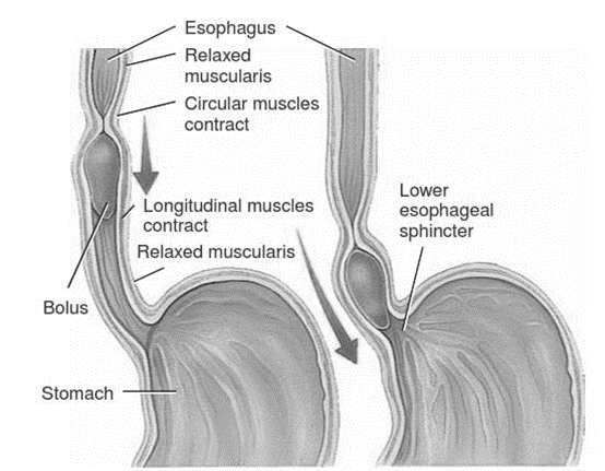
During the esophageal stage of deglutition food moves
through the esophagus into the stomach via peristalsis