Post Views: 130
The Muscular System
The Muscular System
Muscles
Muscles make up almost half the weight of the body
there are 650 different muscles in the human body
the size of your muscles is influenced by how much you use them. this is why speed skaters have large leg muscles
Individual elongated muscle cells can be up to 12 inches, or 30 centimetres, in length
at about the age of 40, the number and diameter of muscle fibers begin to decrease, and by age 80, up to 50 percent of the muscle mass may be lost. exercise and good nutrition help to minimize this loss.
muscle is a general term for all contractile tissue.
Functions of Muscular Tissue
Like nervous tissue, muscles are excitable or “irritable”
they can respond to a stimulus
Unlike nerves, however, muscles are also:
Contractible (they can shorten in length)
Extensible (they can extend or stretch)
Elastic (they can return to their original shape)
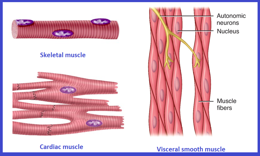
Types of Muscular Tissue
|
Muscle
|
Location
|
Function
|
Appearance
|
Control
|
|
Skeletal
|
skeleton
|
movement,
heat, posture
|
striated, multi-nucleated (eccentric), fibers
parallel
|
voluntary
|
|
Cardiac
|
heart
|
pump blood
continuously
|
striated, one central nucleus
|
involuntary
|
|
Visceral
(smooth
muscle)
|
G.I. tract,
uterus, eye, blood vessels
|
Peristalsis,
blood pressure, pupil size, erects hairs
|
no
striations, one central
nucleus
|
involuntary
|
The Muscular System
Skeletal muscle is the only organ of the muscular system
Skeletal muscle is composed of skeletal muscle tissue and also contains nervous tissue, blood vessels and connective tissue
Half of the body’s weight is muscle tissue
Skeletal muscle = 40% in males, 32% in females
Cardiac muscle = 10%
Skeletal muscle fibers (cells) are arranged into bundles called fascicles
Fascicles are bound by connective tissue
Connective Tissue Coverings
Deep fascia – Surrounds entire skeletal muscle and extends beyond its length
Surrounds an individual skeletal muscle, separating it from other muscles
Fascia may extend beyond the ends of the muscle to become a tendon
Fascia may connect muscle to muscle and is called an aponeurosis
Epimysium – Closely surrounds skeletal muscle, binds fascicles together
Perimysium – Surrounds each fascicle
Endomysium – Surrounds each muscle fiber (cell)
Characteristics of Skeletal Muscle Tissue
Long, thin contractile fibers that are Striated
Striations due to arrangement of thick and thin filaments
Under voluntary control
Attached to the bones of the skeleton by tendons
Allow for movement, facial expressions, breathing, swallowing, writing, talking and singing, posture, heat production, joint stability
Skeletal Muscle Arrangement
A single muscle cell is a muscle fiber
Fibers are made up of myofibrils
Myofibrils are made up of thick and thin filaments
Sarcolemma – muscle cell membrane
Sarcoplasm – muscle cell cytoplasm
Skeletal Muscle Cells
Myofibrils are striated
Striations due to arrangement of thick and thin filaments
Seen as alternating areas of light and dark bands
The length of each myofibril is divided into repeating units called sarcomeres
A sarcomere is the functional unit of skeletal muscle
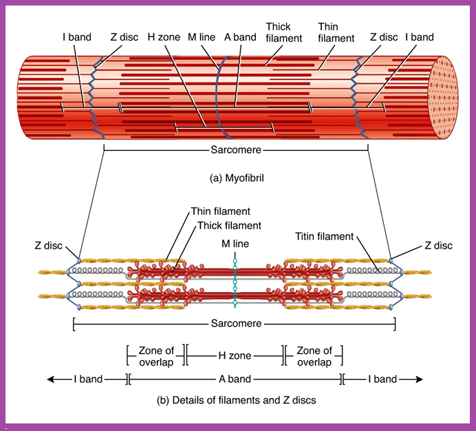
Sarcomere Arrangement
Sarcomere Structure
Sarcomere exists from Z-line to Z-line
A-Band is dark middle band- Overlapping think and thin filaments
I-Band – ends of A-Band, thin filaments only
Z-line is in the middle if the I-Band
Myosin filaments are held to the Z-line by titin proteins
Microscopic anatomy of a skeletal muscle fiber
Thick Filament Structure
Composed of many myosin molecules
Each myosin molecule has a tail region and 2 globular heads (crossbridges)
Thin Filament Structure
Composed of actin protein
2 strands of globular actin molecules twisted into a helix
Actin filaments have binding sites for myosin cross bridges
Tropomyosin protein spirals around actin helix
Troponin protein (3 subunits) is attached to actin and holds tropomyosin in place
Call this the troponin-tropomyosin complex
Specialized Organelles of Skeletal Muscle
Sarcoplasmic Reticulum (SR) – a type of ER
Surrounds each myofibril, running parallel to it
Stores calcium, when stimulated, calcium diffuses into sarcoplasm
Transverse Tubules (TT)
Extends into sarcoplasm as invaginations continuous with sarcolemma
T tubules run between cisternae of SR
Filled with extracellular fluid
Cisternae of SR and TT form a triad near where thick and thin filaments overlap
Also, Visit:
B. Pharma Notes | B. Pharma Notes | Study material Bachelor of Pharmacy pdf
B. Pharma Handwritten Notes
B. Pharma PDF Books
B. Pharma Lab Manual
D. Pharma Lab Manual
B. Pharma 8th Semester Previous Year Question Paper
D. Pharma Notes