Skeletal Muscle Contraction
Motor Neuron
A nerve cell that innervates skeletal muscle tissue
Dendrite –
Receives information
Axon –
Transmits information,
Has vesicles containing neurotransmitters that will stimulate or inhibit muscle contraction
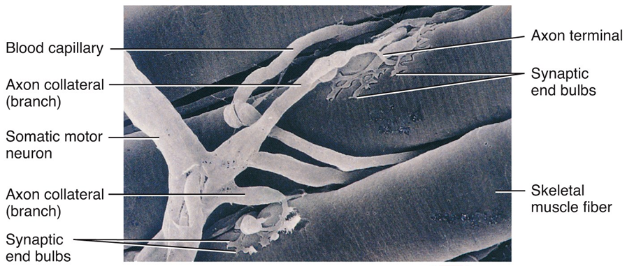
Neuromuscular Junction –
The site where a branch of motor neuron (motor nerve ending) comes in contact with sarcolemma of skeletal muscle fiber
A type of synapse
Neuromuscular Junction
Motor end-plate
Sarcolemma of muscle fiber directly beneath motor nerve ending
Contains an abundance of mitochondria and nuclei
Synapse
The junction between the axonal end of one neuron and the dendrite of another neuron OR membrane of another cell type
Synaptic cleft – Separation that exists between the axonal ending of the motor nerve and the sarcolemma of the muscle cell fiber
The axon ending contains vesicles of neurotransmitter
Neurotransmitter
Chemical substances released from vesicles in the motor nerve ending (axonal ending)
Acetylcholine (Ach) is the neurotransmitter released by motor neurons
When stimulated by a nerve impulse, Ach is released, travels across the synaptic cleft, and binds receptors on the motor end plate
Stimulates contraction
Sliding Filament Theory
A sarcomere is the functional unit of skeletal muscle
When skeletal muscle contracts, sarcomeres shorten
This is described by the sliding filament theory
Sarcomeres shorten because thick and thin filaments slide past one another
Thin filaments move toward the center of the sarcomere from both ends
Physiology of Skeletal Muscle Contraction: Neurotransmitter Release
Motor impulse is initiated in the brain
Travels through the brain and spinal cord to a motor nerve ending
Motor nerve endings (axons) depolarize
Calcium enters the axonal endings
Calcium causes the release of acetylcholine into the neuromuscular junction (synaptic cleft)
Physiology of Skeletal Muscle Contraction: Depolarization
ACh binds receptors on the motor end plate
Depolarizes skeletal muscle fibers
Reverses charge of the membrane
The impulse travels through transverse tubules to reach all of the muscle fibers
Muscle depolarization causes the release of calcium from the sarcoplasmic reticulum into the sarcoplasm
Physiology of Skeletal Muscle Contraction: Power Stroke
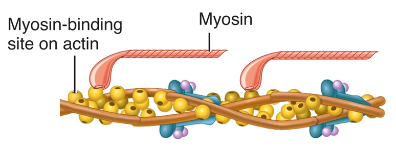
Calcium binds troponin (which is attached to tropomyosin
Moves tropomyosin from the myosin binding sites on actin
Myosin crossbridges (heads) bind to actin
ATP hydrolysis supplies energy
Actin is pulled inward towards the center of the sarcomere = POWER STROKE
Sarcomeres shorten as muscle contracts
The Sliding-Filament Mechanism
With the exposure of the myosin binding sites on actin (the thin filaments)—in the presence of Ca2+ and ATP—the thick and thin filaments “slide” on one another and the sarcomere is shortened
Muscle Relaxation Mechanism
1. Acetylcholinesterase present in the NMJ destroys ACh (preventing continual stimulation)
2. Calcium ions are transported from the sarcoplasm back into the SR
3. Linkages between myosin and actin are broken – Requires ATP binding
THEN: The muscle fiber relaxes
Contraction in the Sarcomere
A band stays the same
I band gets smaller
H zone gets smaller
Sarcomere shortens
Energy for Contraction
Muscle cells require huge amounts of ATP energy to power contraction
The cells have only a very small store of ATP
Three pathways supply ATP to power muscle contraction
ATP initially supplied from cellular respiration
If ATP is abundant, is converted to creatine phosphate and stored in skeletal muscles
When ATP is low, creatine phosphate supplies phosphate to ADP making ATP
CP & ATP stores only good for about a 10-second maximal contraction
ATP must then come from cellular respiration or glycolysis
Oxygen & Muscle Contraction
Myoglobin of muscle (similar to hemoglobin) binds to and stores oxygen
Supplies O2 needed to make ATP for contraction
Exercise and Skeletal Muscles
Prolonged, moderate exercise
ATP supplied through cellular respiration
Once glycogen stores are depleted in muscle, glucose and fatty acid deliveries from blood are used as fuel source
Intense, strenuous exercise
Muscles exceed the capacity of respiratory and cardiovascular systems to deliver oxygen for contraction
ATP supplied anaerobically through glycolysis
Pyruvate is converted to lactic acid
Lactic acid builds up in muscles
Causes muscles to fatigue
Exercise and Contraction
In intense strenuous activity, oxygen can be depleted
Contraction of skeletal muscles decreases blood delivery to muscles
Nutrient and O2 levels in contracting muscles decrease
Oxygen Debt
Amount of oxygen needed by liver cells to use the accumulated lactic acid to produce glucose
Oxygen not available
Glycolysis continues
Pyruvic acid converted to lactic acid
The liver converts lactic acid to glucose
Also the amount of oxygen needed to replace O2 levels in skeletal muscle to pre-exercise levels
Heat Production
Cellular respiration is only about 40% efficient
About 60% of the energy found in glucose is lost as heat during cellular respiration
Muscle contraction generates heat because muscles use large amounts of nutrients to make ATP, generating large amounts of heat
Heat is used to maintain body temperature
Glycogenolysis
Glycogenolysis is the breakdown of glycogen into glucose molecules
Epinephrine can trigger this pathway too
Depends upon the presence of an enzyme – glycogen phosphorylase
McArdle’s Disease
Absence of the muscle glycogen phosphorylase enzyme
Individuals must rely on blood-transported fuels
Fatty acids, protein, and glucose from the liver
These reserves take 5-10 minutes to arrive in the mitochondria
Muscles stop functioning until these fuels arrive
Autosomal Recessive Disorder
Symptoms of McArdle’s Disease
Premature muscle fatigue and weakness and pain during exercise
Muscles can become injured during exercise
The Muscular System & Skeletal Muscle Contraction Notes PDF

Also, Visit: Pharmacology Notes