Virus Classification, Structure, Function and Methods for Cultivation

Virus
Virus- infectious agent of small size and simple composition that can multiply only in living cells of animals, plants, or bacteria. The name is from a Latin word meaning “slimy liquid” or “poison.” an extremely tiny parasite that can only reproduce if it is within a living being
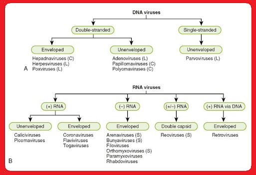
Classification of viruses
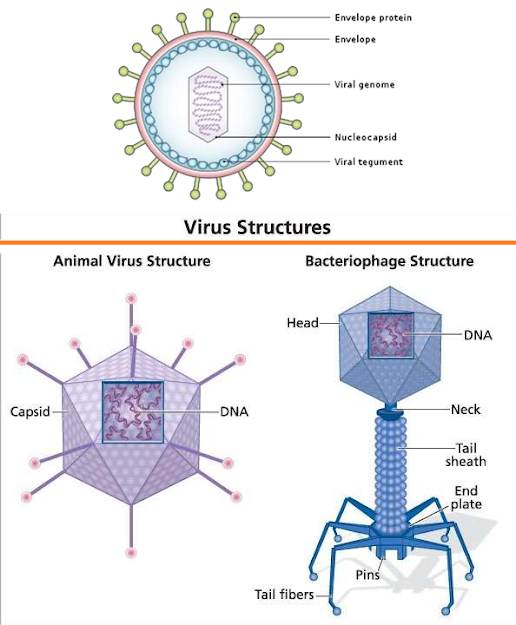
Virus Structure
Morphology of Virus
Viruses are grouped based on size and shape, chemical composition and structure of the
genome, and mode of replication. Helical morphology is seen in nucleocapsids of many filamentous and pleomorphic viruses.
Helical nucleocapsids consist of a helical array of capsid proteins (protomers) wrapped around a helical filament of nucleic acid. Icosahedral morphology is characteristic of the nucleocapsids of
many “spherical” viruses.
The number and arrangement of the capsomeres (morphologic subunits of the icosahedron) are
useful in identification and classification. Many viruses also have an outer envelope
Structure and Function of Virus
1. Viruses are inert outside the host cell. Small viruses, e.g., polio and tobacco mosaic virus, can even be crystallized.
2. Viruses are unable to generate energy. As obligate intracellular parasites, during replication, they fully depend on the complicated biochemical machinery of eukaryotic or prokaryotic cells. The main purpose of a virus is to deliver its genome into the host cell to allow its expression (transcription and translation) by the host cell.
3. A fully assembled infectious virus is called a virion. The simplest virions consist of two basic components: nucleic acid (single- or double-stranded RNA or DNA) and a protein coat, the capsid, which functions as a shell to protect the viral genome from nucleases and which during infection attaches the virion to specific receptors exposed on the prospective
4. host cell. Capsid proteins are coded for by the virus genome. Because of its limited size the genome codes for only a few structural proteins (besides non-structural regulatory proteins involved in virus replication).
5. Capsids are formed as single or double protein shells and consist of only one or a few structural protein species. Therefore, multiple protein copies must self-assemble to form the continuous three-dimensional capsid structure. Self-assembly of virus
6. capsids follows two basic patterns: helical symmetry, in which the protein subunits and the nucleic acid are arranged in a helix, and icosahedral symmetry, in which the protein subunits assemble into a symmetric shell that covers the nucleic acid-containing core.
Viral Replication
Steps in Viral Replication
Attachment. This is the first step in viral replication. Surface proteins of the virus interact with specific receptors on the target cell surface. These may be specialized proteins with limited distribution or molecules that are more widely distributed on tissues throughout the body. The presence of a virus-specific receptor is necessary but not sufficient for viruses to infect cells and complete the replicative cycle.
Penetration. Enveloped viruses (e.g., HIV, influenza virus) penetrate cells through fusion of the viral envelope with the host cell membrane. Non-enveloped viruses penetrate cells by translocation of the virion across the host cell membrane or receptor mediated endocytosis of the virion in clathrin coated pits with accumulation of viruses in cytoplasmic vesicles.
Uncoating (disassembly). A complex process which differs by taxonomic class and is not fully understood for many agents. This process makes the nucleic acid available for transcription to permit multiplication of the virus.
Transcription and Translation. The key to understanding the genomic expression of viruses is noting the fact that viruses must use host cellular machinery to replicate and make functional and structural proteins. Strategies for genomic expression for different taxonomic groupings of viruses are described below (section II).
Assembly and Release. The process of virion assembly involve bringing together newly formed viral nucleic acid and the structural proteins to form the nucleocapsid of the virus. There are basically three strategies that viruses employ:
1. Nonenveloped viruses exhibit full maturation in the cytoplasm (e.g., picornaviruses) or the nucleus (e.g.,adenoviruses) with disintegration of the cell and release of virions.
2. For enveloped viruses, including the (-) strand RNA viruses, the (+) strand togaviruses and the retroviruses, final maturation of the virion takes place as the virion exits the cell.
Viral proteins are inserted into the host cell membrane. Nucleocapsids bind to the regions of the host cell membranes with these inserted proteins and bud into the extracellular space. Further cleavage and maturation of proteins may occur after viral 2 extrusion to impart full infectivity on the virion. Viruses in this group differ in their degree of cytolytic activity.
3. Herpesviruses, which are enveloped viruses, assemble their nucleocapsids in the nuclei of infected cells and mature at the inner lamella of the nuclear membrane. Virions accumulate in this region, in the endoplasmic reticulum and in vesicles protected from the cytoplasm. Release of virions from the cell surface is associated with cytolysis.
Methods for Cultivation of Virus
Since the viruses are obligate intracellular parasites, they cannot be grown on any inanimate
culture medium. Viruses can be cultivated within suitable hosts, such as a living cell. Generally three
methods are employed for the virus cultivation.
1. Inoculation of virus into animals.
2. Inoculation of virus into embryonated eggs.
3. Tissue culture- refer to tissue culture topic- Organ cultue /Explant culture/ cell culture
Inoculation of Virus in Animals
Laboratory animals are widely used for routine cultivation of virus; they play an esse ntial role instudies of viral pathogenesis.
Live animals such as monkeys, mice, rabbits, guinea pigs, ferrets are widely used for cultivating virus.
Monkeys were used for the isolation of Poliovirus.
But due to their risk to handlers, monkeys find only limited applications in Virology.
Mice are the most widely employed animals in virology.
The different routes of inoculation in mice are intracerebral, subcutaneous, intraperitoneal or intranasal.
After the animal is inoculated with the virus suspension, the animal is observed for signs of disease, visible lesions or is killed so that infected tissues can be examined for virus.
Advantages:
1. Animal inoculation may be used as diagnostic procedure for identifying and isolating a virus
from a clinical specimen.
2. Mice provide a reliable model for studying viral replication.
3. Gives unique insight into viral pathogenesis and host virus relation.
4. Used for the study of immune responses, epidemiology and oncogenesis.
Disadvantages:
Expensive and difficulties in maintenance of animals.
Inoculation of Virus into Embryonated eggs
Prior to the advent of cell culture, animal viruses could be propagated only on whole animals or embryonated chicken eggs.
Good pasture in 1931first used the embryonated hen’s egg for the cultivation of virus.
The process of cultivation of viruses in embryonated eggs depends on the type of egg which is used.
The egg used for cultivation must be sterile and the shell should be intact and healthy.
A hole is drilled in the shell of the embryonated egg, and a viral suspension or suspected virus- containing tissue is injected into the fluid of the egg.
Viral growth and multiplication in the egg embryo is indicated by the death of the embryo, by embryo cell damage, or by the formation of typical pocks or lesions on the egg membranes. An embryonated egg offers various sites for the cultivation of viruses
The different sites of viral inoculation in embryonated eggs are:
Chorioallantoic membrane(CAM).
Amniotic Cavity.
Allantoic Cavity.
Yolk sac.
Embryo.
Air sac.
Amniotic Cavity.
Allantoic Cavity.
Yolk sac.
Embryo.
Air sac.
Frequently Asked Questions (FAQs)
- Are all viruses harmful to humans?
- While some viruses can cause diseases, not all are harmful. Some viruses are essential for various biological processes.
- How do viruses evolve?
- Viruses evolve through mutations, allowing them to adapt to changes in their environment.
- Can viruses be beneficial?
- Yes, certain viruses play crucial roles in processes such as genetic diversity and the evolution of species.
- What is the role of vaccination in preventing viral infections?
- Vaccination stimulates the immune system to recognize and fight specific viruses, preventing infections and promoting herd immunity.
- How can the public contribute to virus research?
- Staying informed, supporting scientific initiatives, and participating in awareness campaigns are ways the public can contribute to virus research.
