Human Eye
Objectives
At the end of this lecture, student will be able to
• Describe the structural components of eye ball
• Explain the accessory structures of eye ball
• Distinguish between the structural components and the
accessory structures of eye ball
• Describe the interior of the eye ball
• Explain image formation
• Explain the physiology of vision
• Distinguish the changes occurring during light and dark
adaptation
• Explain the processing of visual signals in retina
THE EYE
• Organ of the sense of sight
• Responsible for the detection of visible light (400-700nm)
• Location – In the orbital cavity; supplied by optic nerve
Accessory Structures of the Eye
• The eyelids
• Eyelashes
• Eyebrows
• The lacrimal apparatus
• Extrinsic eye muscles
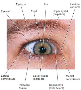
Eyelids
• Upper and lower eyelids, or palpebrae
• Shade the eyes during sleep
• Protect eyes from excessive light and foreign objects
• Spread lubricating secretions over the eyeballs
• Upper eyelid more movable than the lower
• Contains in its superior region the levator palpebrae
superioris muscle
• Palpebral fissure – space between the upper and lower eyelids that exposes the eyeball
• Angles known as lateral commissure & medial commissure
• Lateral commissure- narrower and closer to the temporal bone
• Medial commissure- broader and nearer to the nasal bone
• A small, reddish elevation, the lacrimal caruncle contains
sebaceous (oil) glands and sudoriferous (sweat) glands
From superficial to deep, each eyelid consist of
• Epidermis
• Dermis
• Subcutaneous tissue
• Fibers of the orbicularis oculi muscle
• A tarsal plate – thick fold of connective tissue; supports eyelid
• Tarsal glands – Modified sebaceous glands (Meibomian glands)
• Conjunctiva – Thin, protective mucous membrane composed of non-keratinized stratified columnar epithelium
• Palpebral conjunctiva – lines the inner aspect of the eyelids
• Bulbar conjunctiva – passes from the eyelids onto the surface of the eye ball covers the sclera

Eyelashes and Eyebrows
• Eyelashes – Project from the border of each eyelid
• Eyebrows – Arch transversely above the upper eyelids
• Help protect the eyeballs from – foreign objects
– Perspiration
– The direct rays of the sun
• Sebaceous ciliary glands – Sebaceous glands at the base of the hair follicles of the eyelashes
• Release a lubricating fluid into the follicles
• Infection of these glands is called a sty
The Lacrimal Apparatus
A group of structures that produces and drains lacrimal fluid or tears

• Lacrimal glands – supplied by parasympathetic fibers, facial (VII) nerves
• Lacrimal fluid – a watery solution has salts, some mucus, lysozyme, a protective bactericidal enzyme
• Lacrimation – a protective mechanism
– The tears dilute and wash away the irritating substance
• Crying – Excessive lacrimal fluid production by lacrimal glands in response to parasympathetic stimulation
Extrinsic Eye Muscles
• Extend from the walls of the bony orbit to the sclera (white) of the eye
• Surrounded in the orbit by periorbital fat
• Capable of moving the eye in almost any direction
Six extrinsic eye muscles move each eye
• Superior rectus
• Inferior rectus
• Lateral rectus
• Medial rectus
• Superior oblique
• Inferior oblique
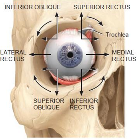
Extrinsic eye muscles that move the eyeballs and upper eyelid

• Supplied by cranial nerves III, IV, or VI
• Extrinsic eye muscles move the eyeball laterally, medially, superiorly, and inferiorly
• Oblique muscles preserve rotational stability of the eyeball
• Neural circuits in the brain stem and cerebellum coordinate and synchronize movements of eye
Accessory structures of the eye

Anatomy of the Eyeball
• Adult eyeball – about 2.5 cm in diameter
• Only anterior one-sixth exposed
• Remainder protected by the orbit
Wall of the eyeball consists of three layers:
(1) Fibrous tunic (sclera & cornea)
(2) Vascular tunic (choroid, ciliary body, and iris), and
(3) Retina
(1) Fibrous Tunic
• Superficial layer of the eye
• Consists of –
(a) The anterior cornea
(b) Posterior sclera
(a) The cornea
• Transparent coat; covers the colored iris
• Helps focus light onto the retina as it is curved
• Outer surface – non-keratinized stratified squamous epithelium
• Middle coat – collagen fibers and fibroblasts,
• Inner surface – simple squamous epithelium

(b) The sclera
• The “white” of the eye
• Covers the entire eyeball except the cornea
• Gives shape and rigidity to the eyeball
• Protects its inner parts
• Serves as a site of attachment for the extrinsic eye
muscles
• At the junction of the sclera and cornea is an opening known as the scleral venous sinus (canal of Schlemm)
• A fluid called aqueous humor drains into this sinus
(2) Vascular Tunic/Uvea
• Middle layer of the eye ball
• Composed of three parts:
(a) Choroid
(b) Ciliary body
(c) Iris

(a) Choroid
• Highly vascularized
• Provide nutrients to the posterior surface of the retina
• Contains melanocytes, produce the pigment melanin (dark brown)
• Melanin in the choroid absorbs stray light rays
• Prevent reflection and scattering of light within the
eyeball
(b) Ciliary body
• Anterior portion of the vascular tunic, the choroid becomes the ciliary body
Ciliary body consists of:
Ciliary processes
• Protrusions or folds on the internal surface of the ciliary body
• Extentions from ciliary process, zonular fibres (suspensory
ligaments); attach to lens
Ciliary muscle
• A circular band of smooth muscle
• Changes the tightness of the zonular fibers lters the shape of the lens dapt lens for near or far vision
(c) Iris
• Iris (= rainbow), the colored portion of the eyeball
• Suspended between the cornea and the lens
• Consists of melanocytes and circular (sphincter pupillae)
and radial smooth muscle fibers (dilator pupillae)
• Amount of melanin in the iris determines the eye color
• Brown to black – large amount of melanin
• Blue – low melanin
• Green – moderate melanin concentration
• Iris regulate the amount of light entering the eyeball through the pupil
• Autonomic reflexes regulate pupil diameter in response to
light levels

(3) Retina
• Lines the posterior three-quarter of the eyeball
• Is the beginning of the visual pathway

• Optic disc – site, optic (II) nerve exits the eyeball
• Central retinal artery, a branch of the ophthalmic artery, and the central retinal vein are bundled with optic disc
Transverse section of posterior eyeball at optic disc

Retina consists of a pigmented layer and a neural layer
|
Pigmented |
Neural |
|
• Sheet of melanin-containing epithelial cells • Located between the choroid and the neural part of the retina • Melanin – also helps to absorb stray light rays |
• Multilayered outgrowth of the brain • Processes visual data extensively before sending nerve impulses
|
Three distinct layers of retinal neurons
• The photoreceptor layer
• The bipolar cell layer
• The ganglion cell layer
• Separated by two zones, the outer and inner synaptic
layers
• Two other types of cells present in the bipolar cell layer
of the retina are called horizontal cells and amacrine cells
Photoreceptors
Specialized cells that begin the process of conversion of
light rays to nerve impulses
Two types of photoreceptors: rods and cones
Rods – allow to see in dim light, such as moonlight
– do not provide color vision, only black and white
Cones – stimulated by brighter lights
– produce color vision
• Three types of cones
Blue cones- sensitive to blue light
Green cones-sensitive to green light
Red cones –sensitive to red light
Microscopic structure of the retina

• Optic disc or blind spot, contains no rods or cones
• We cannot see an image that strikes the blind spot
• Macula lutea or yellow spot is in the exact center of the
posterior portion of the retina, at the visual axis of the eye
• Fovea centralis – Small depression in the center of the macula lutea
– Contains only cones
– Area of highest visual acuity or resolution (sharpness of vision)
Lens
• Behind the pupil and iris, within the cavity of the eyeball
• In the cells of the lens, proteins called crystallins, arranged like the layers of an onion
• Make up the refractive media of the lens
• Transparent and lacks blood vessels
• Enclosed by a clear connective tissue capsule
• Held in position by encircling zonular fibers, which attach
to the ciliary processes
• Lens helps focus images on the retina to facilitate clear
vision
Anatomy of the eyeball

Interior of the Eyeball
Lens divides the interior of the eyeball into two cavities: Anterior cavity and Vitreous chamber
1. Anterior cavity- space anterior to the lens
Consists of two chambers
• Anterior chamber – between the cornea and the iris
• Posterior chamber – behind iris and in front of zonular
fibers and lens
• Both chambers of the anterior cavity are filled with
aqueous humor
• Transparent watery fluid that nourishes the lens and cornea
• Completely replaced about every 90minutes

2. Vitreous chamber
• Larger posterior cavity of the eyeball
• Lies between the lens and the retina
Vitreous body – a transparent jellylike substance
• Holds the retina flush against the choroid
• Gives the retina an even surface for the reception of
clear images
• Contains phagocytic cells, remove debris
• Keep the eye clear for unobstructed vision

Intraocular pressure – pressure in the eye
• Produced mainly by aqueous humor and partly by vitreous body
• Normally it is about 16 mmHg
• Maintains the shape of the eyeball
• Prevents it from collapsing
Interior of the Eyeball

Image Formation
Image formation involves 3 processes
(1) The refraction or bending of light by the lens and cornea
(2) Accommodation, the change in shape of the lens
(3) Constriction or narrowing of the pupil
Refraction of light rays
Refraction is the bending of light rays at the junction of two transparent substances with different densities

Accommodation
• Increase in the curvature of the lens for near vision, Accommodation
• Viewing a close object, the ciliary muscle contracts, which pulls the ciliary process and choroid forward toward the lens, become more spherical (more convex)
• Near point of vision – minimum distance from the eye that an object can be clearly focused with maximum accommodation

• Distance about 10 cm in a young adult
Convergence
• In humans, both eyes focus on only one set of objects—a characteristic called binocular vision
• In convergence, the eyeballs move medially so they are
both directed toward an object being viewed
Physiology of Vision
Photoreceptors and Photo pigments
• Rods and cones have different appearance of the outer
segment
• Rods – cylindrical/ rod-shaped, plasma membrane (PM) form discs
• Cones – tapered/ cone-shaped, PM folded back and forth
• Photo pigments, integral proteins in the plasma membrane
of the outer segment
• Inner segment – contains the cell nucleus, Golgi complex,
and many mitochondria
Structure of rod and cone photoreceptors
• Inner segments contain the metabolic machinery for
synthesis of photo pigments and production of ATP
• Transduction of light energy into a receptor potential
occurs in the outer segments of rods and cones

Photo pigment
• A colored protein, undergoes structural changes when it
absorbs light, in the outer segment of a photoreceptor
• Light absorption initiates production of a receptor potential
• Photo pigment in rods – rhodopsin; in cones – 3 types
• Photo pigments associated with vision contain two parts:
– A glycoprotein, opsin
– A derivative of vitamin A, retinal
• Retinal – light-absorbing part of all visual photo pigments
• In humans, 4 different opsins; 3 in cones; 1 in rods
The cyclical bleaching and regeneration of photopigment
• Blue arrows indicate bleaching steps
• Black arrows indicate regeneration steps

Light and Dark Adaptation
Light adaptation— when emerging from a dark surrounding to sunshine
• Visual system adjusts in seconds to the brighter environment by decreasing its sensitivity
• As the light level increases, more and more photo pigment
bleached
• Other photo pigments regenerate
• Regeneration of rhodopsin is insignificant
• Rods contribute little to daylight vision
• Cone photo pigments regenerate rapidly
Dark adaptation- when entering a dark room
• Sensitivity increases slowly over many minutes
• Full regeneration of cone photo pigments occurs during the
first 8 min of dark adaptation
• Rhodopsin regenerates more slowly
• Our visual sensitivity increases until even a single photon (the smallest unit of light) can be detected
• Threshold flashes appear gray-white, regardless of their
color
Release of Neurotransmitter by Photoreceptors In darkness

The Visual Pathway
Processing of Visual Input in the Retina
• Receptor potentials arise in the outer segments of rods
and cones
• Neurotransmitter molecules released by rods and cones
• Induce local graded potentials in both bipolar cells and
horizontal cells
• Horizontal cells transmit inhibitory signals to bipolar cells
• Bipolar or amacrine cells transmit excitatory signals to
ganglion cells
• Ganglion cells depolarize and initiate nerve impulses
Brain Pathway and Visual Fields
• From the thalamus, impulses cerebral cortex (occipital
lobe)
• Axon collaterals of retinal ganglion cells extend to the midbrain and hypothalamus

• Everything that can be seen by one eye – Eye’s visual
field
• We have binocular vision due to the large region where the
visual fields of the two eyes overlap—the binocular visual field
Visual field of each eye is divided into two regions:
a) The nasal or central half
b) The temporal or peripheral half
• Light rays from an object in the nasal half of the visual
field fall on the temporal half of the retina, and vice versa
• Visual information from the right half of each visual
field is conveyed to the left side of the brain, and vice versa
Transverse section through eyeballs and brain

The visual pathway

Summary
• Eye is the organ of the sense of sight
• Eye is situated in the orbital cavity supplied by optic
nerve
• Accessory structure of eye ball – eyelids, Eyelashes, Eyebrows, Lacrimal apparatus, extrinsic eye muscles
• Eyelashes & eyebrows protects the eye ball
• Lacrimal apparatus produces and drains lacrimal fluid or
tears
• Extrinsic eye muscle moving the eye in almost any direction
• Wall of the eyeball consists of – fibrous tunic (sclera and cornea), vascular tunic (choroid, ciliary body, and iris), and retina
• Lens helps focus images on the retina to facilitate clear
vision
• Lens divides the interior of the eyeball into anterior cavity and the vitreous chamber
• Anterior cavity consists of anterior and posterior chamber
• Image formation involves – refraction of light, accommodation and convergence
• Cyclic bleaching and regeneration of photo pigments helps
in vision
• Light adaptation occur when emerging from a dark
surrounding to sunshine
• Dark adaptation occur when entering a dark surrounding
• Visual field of each eye consists of nasal region and
temporal region
Also, Visit:
B. Pharma Notes | B. Pharma Notes | Study material Bachelor of Pharmacy pdf