Human Skin
Objectives
At the end of this lecture, student will be able to
• Describe the layers of the epidermis and the cells that
compose them
• Describe various accessory structures of the skin
• Distinguish between the accessory structures and the main
components of skin
• Explain the functions of skin
Content
• Skin
– Layers of epidermis
– Accessory structures
– Function
The Skin
• Also known as the cutaneous membrane or integument
• Covers the external surface of the body
• Largest organ of the body in both surface area and weight
Structurally, the skin consists of two main parts
1. Epidermis – superficial, thin epithelial tissue portion
2. Dermis – deep, thicker connective tissue portion
Subcutaneous layer (Hypodermis), attaches the dermis to underlying fascia
Functions of subcutaneous layer
Subcutaneous layer serves
• As a storage depot for fat
• Contains large blood vessels that supply to the skin
• Also contains nerve ending called pacinian (lamellated) corpuscles that are sensitive to pressure
Components of the integumentary system
Epidermis
• Composed of keratinized stratified squamous epithelium
• Contains four principal types of cells
Keratinocytes
Melanocytes
Langerhans cells
Merkel cells
Keratinocytes – 90% of epidermal cells
• Arranged in four or five layers
• Produce the protein keratin
• Protect the skin and underlying tissues from heat, microbes, and chemicals
Melanocytes
• Develop from the ectoderm of a developing embryo
• Produce the pigment melanin
• Contributes to skin color and absorbs damaging UV light
• Long, slender projections extend between the keratinocytes
• Transfer melanin granules to keratinocyte
Langerhans cells
• Arise from red bone marrow and migrate to the epidermis
• Constitute a small fraction of the epidermal cells
• Participate in immune responses
• Recognize an invading microbe and destroy it
Merkel cells
• Least numerous of the epidermal cell
• Deepest layer of the epidermis
• Contact the flattened process of a sensory neuron, a merkel (tactile) disc
• Detect touch sensations
Layer of Epidermis
|
4 strata layers in most region
|
5 layers where friction is greatest (fingertips, palms, & Soles) |
|
Stratum basale Stratum spinosum Stratum granulosum Thin stratum corneum (Thin skin) |
Stratum basale Stratum spinosum Stratum granulosum Thick stratum corneum (Thick skin) Stratum lucidum |
Stratum basale
• Deepest layer of the epidermis
• Single row of cuboidal or columnar tinocytes
• Stem cells that undergo cell division to continually produce new keratinocytes
• Contain scattered tono filament (intemediate filaments)
• Melanocytes and merkel cells associated with merkel discs are scattered among the keratinocytes
Stratum spinosum
• Eight to ten rows of many-sided keratinocytes
• Bundles of tono filaments; includes arm like processes of melanocytes and langerhans cells
• Arrangement provides both strength and flexibility to the skin
Stratum granulosum
• Three to five rows of flattened keratinocytes
• Organelles are beginning to degenerate
• Cells contain the protein keratohyalin, converts tono filaments into keratin
• Lamellar granules, which release a lipid-rich, water-repellent secretion
Stratum lucidum
• Present only in skin of fingertips, palms, and soles
• Consists of 3-5 rows of clear, flat, dead keratinocytes with large amounts of keratin
Stratum corneum
• Twenty-five to thirty rows of dead, flat keratinocytes that contain mostly keratin
Dermis
• Deeper part of the skin
• Composed of a strong connective tissue containing collagen and elastic fibers
• Fibers has great tensile strength
• Ability to stretch and recoil easily
• Based on its tissue structure, the dermis is divided into
a) A superficial papillary region
b) A deeper reticular region
Papillary region
• The superficial portion of the dermis
• Consists of areolar connective tissue with thin collagen and fine elastic fibers
• Contains dermal ridges that has capillaries, Meissner corpuscles, and free nerve endings
Reticular region
• Deeper portion of the dermis
• Consists of dense irregular connective tissue with bundles of thick collagen and coarse elastic fibers
• Spaces between fibers contain some adipose cells, hair follicles, nerves, sebaceous glands, and sudoriferous glands
Accessory structures of the skin
• Hair
• Skin glands
• Nails—develop from the embryonic epidermis
Hair
• Formed by the down growth of epidermal cells into dermis or subcutaneous tissue, Hair follicles
• At the base of the follicle is a cluster of cells, bulb
• Hair – formed by the multiplication of cells of bulb
• Pushed upward away from the source of nutrition, the cells die and get keratinised
• The part of hair above the skin, Shaft, the remainder is the root
• Arrector pili – little bundles of smooth muscle fibres attached to the hair follicles
• Contraction makes the hair stand erect and raise the skin around the hair causing goose flesh
Skin glands
Exocrine glands are associated with the skin:
• Sebaceous (oil) glands
• Sudoriferous (sweat) glands
• Ceruminous glands
Sebaceous (oil) glands
• Simple, branched acinar glands
• Connected to hair follicles; absent from the palms and soles
• Produce sebum, moistens hairs and waterproofs the skin
• Clogged sebaceous glands may produce acne
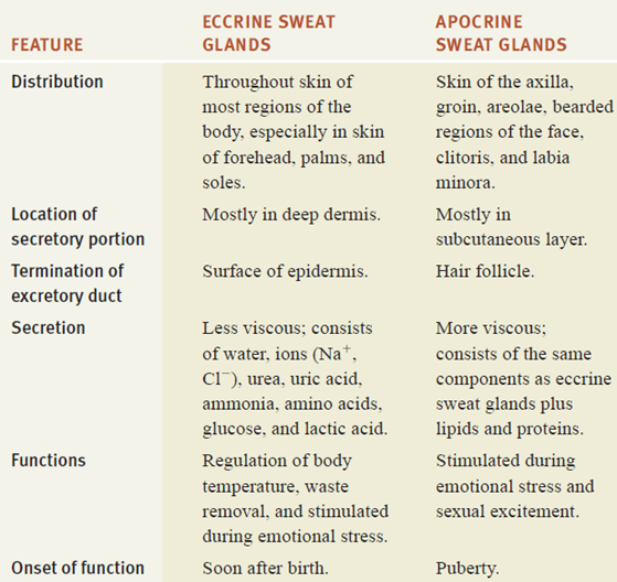
Sudoriferous (sweat) glands
Release sweat, or perspiration, into hair follicles or onto the skin surface through pores
Two types of sweat glands
– Eccrine glands
– Apocrine glands
Ceruminous glands
• Nodified sudoriferous glands
• Secrete cerumen, found in the external auditory canal (ear canal)
Types of sweat glands
Nails
Plates of tightly packed, hard, dead, keratinized epidermal cells
Each nail consists of
• A nail body (plate) – exposed part grown out from the germinative zone of epidermis
• A free edge – part of the nail body that may extend past the distal end of the digit
• A nail root – portion of the nail that is buried in a fold of skin
Nail and its internal details
Functions of the Skin
• Body temperature regulation – liberating sweat at its surface and by adjusting the flow of blood in the dermis
• Blood storage
• Excretion and absorption, and synthesis of vitamin D
• Provides physical, chemical, and biological barriers that help protect the body
• Cutaneous sensations include tactile sensations, thermal sensations, and pain
Summary
• Skin is the largest organ of the body in surface area and
weight
• Principal parts of the skin are the epidermis
(superficial) and dermis (deep)
• Types of cells in the epidermis are keratinocytes,
melanocytes, Langerhans cells, and Merkel cells
• Epidermal ridges provide the basis for fingerprints and
footprints
• The color of skin is due to melanin, carotene, and
hemoglobin
• Accessory structures of the skin—hair, skin glands, and
nails—develop from the embryonic epidermis
• Skin functions include body temperature regulation, blood
storage, protection, sensation, excretion and absorption, and synthesis of
vitamin D


