The Small Intestine
The Small
Intestine
• Most digestion and absorption of nutrients occur
• Length provides a large surface area
• Area is further increased by circular folds, villi, and
microvilli
• Begins at the pyloric sphincter of the stomach
• Eventually opens into the large intestine
• Length – 3 m in a living person and about 6.5m in a
cadaver
Parts of
Small Intestine
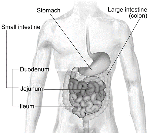
Anatomy of
Small Intestine
• Duodenum – 25 cm
– Shortest region 25 cm
– Retroperitoneal
– Merges with the jejunum
• Jejunum – 1 m
– 1 m long and extends to the ileum
– Jejunum means “empty” – found at death
• Ileum – 2 m
– The final and longest region of the small intestine
– Joins the large intestine at ileocecal sphincter
Functions
of Small Intestine
• Segmentations mix chyme with digestive juices and bring
food into contact with the mucosa for absorption
• Peristalsis propels chyme through the small intestine
• Completes the digestion of carbohydrates, proteins, and
lipids
• Begins and completes the digestion of nucleic acids
• Absorbs about 90% of nutrients and water
Layers of
Small Intestine

Histology
of Small Intestine
• Composed of a layer of epithelium, lamina propria &
muscularis mucosae
• Epithelial layer:
consists of simple columnar epithelium
– Absorptive cells, Goblet cells & Paneth cells
– Enteroendocrine cells: S cells, CCK cells and K cells
• Intestinal glands (crypts of Lieberkühn) and secrete
intestinal juice

Mucosa
• The lamina propria
– Contains areolar connective tissue
– Abundance of mucosa-associated lymphoid tissue (MALT)
– Ileum: Groups of lymphatic nodules – aggregated lymphatic
follicles (Peyer’s patches)
– The muscularis mucosae
– Consists of smooth muscle
Enlarged Villus

Histology
of Small Intestine – Submucosa
• Duodenum contains duodenal (Brunner’s) glands – secrete an
alkaline mucus
• Helps neutralize gastric acid in the chyme
Muscularis
• Consists of two layers of smooth muscle
• The outer, thinner layer contains longitudinal fibers
• The inner, thicker layer contains circular fibers
Serosa
• Except for a major portion of the duodenum, the serosa (or
visceral peritoneum) completely surrounds the small intestine
Special
Features of Small Intestine
• Facilitate the process of digestion and absorption
• The structural features include:
– Circular folds, Villi & Microvilli

Circular
folds or Plicae circulares
• Folds of the mucosa and submucosa
• These permanent ridges – 10 mm long
• Begin near the proximal portion of the duodenum
• End at about the midportion of the ileum
• Enhance absorption by increasing surface area
• Causing the chyme to spiral, rather than move in a
straight line
Villi (Tufts
of Hair)
• Fingerlike projections of the mucosa – 0. 5–1 mm long
• Gives the intestinal mucosa a velvety appearance
• Each villus – covered by epithelium and has a core of
lamina propria

Microvilli
• Projections of the apical (free) membrane of the
absorptive cells
• Each microvillus is a 1 m-long cylindrical, membrane
-covered projection
• Contains a bundle of 20–30 actin filaments
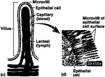
• Greatly increase the surface area
• Viewed through a light microscope as fuzzy line – brush
border
• Brush border contains several brush-border enzymes – have
digestive functions
• 200 million microvilli per square millimeter of small
intestine
Role of
Intestinal Juice and Brush Border Enzyme
Intestinal
juice
• Clear yellow fluid
• Contains water and mucus and is slightly alkaline (ph 7.6)
• Together, pancreatic and intestinal juices provide a
liquid medium
• Aids the absorption of substances from chyme
Brush-border
Enzymes
• The absorptive cells of the small intestine synthesize
several digestive enzymes brush-border enzymes
• Insert them in the plasma membrane of the microvilli
Carbohydrate
digesting enzyme
• Dextrinase – dextrins into glucose
• Maltase – Maltose to glucose
• Sucrase – Sucrose to glucose and fructose
• Lactase – Lactose to glucose and galactose
Protein-digesting
enzymes – Peptidases
• Aminopeptidases – Break off amino acids at the amino ends
of peptides
• Dipeptidases – Split dipeptides into aminoacids
Nucleotide-digesting
enzymes
• Nucleosidases and Phosphatases – Nucleotides to pentoses
and nitrogenous bases
Mechanical
Digestion in Small Intestines
• The two types of movements of the small intestine:
– Migrating Motility Complexes – type of peristalsis called
governed by the myenteric plexus
– Segmentations

• Segmentations
– Localized, mixing contractions
– Occur in portions of intestine distended by a large volume
of chyme
– Segmentations mix chyme with the digestive juices
– Bring the particles of food into contact with the mucosa
for absorption
– They do not push the intestinal contents along the tract
– Duodenum – 12 times per minute
– Progressively slow to – 8 times per minute in the ileum
MMC
• Less distension of the small intestine, segmentation stops
and peristalsis begins
• Begins in the lower portion of the stomach
• Pushes chyme forward along a short stretch of small
intestine
• Slowly migrates down the small intestine, reaching the end
of the ileum in 90–120 minutes
• Altogether, chyme remains in the small intestine for 3–5
hours
Chemical
Digestion in Small Intestines
• In the mouth
salivary amylase converts starch (a polysaccharide) to:
• Maltose (a disaccharide)
• Maltotriose (a trisaccharide)
• Dextrins (short-chain, branched fragments of starch with
5– 10 glucose units)
• Lingual and gastric
lipases convert some triglycerides into:
– Fatty acids
– Diglycerides
– monoglycerides
• In the stomach,
pepsin converts proteins to:
– Peptides (small fragments of proteins)
• Thus, chyme entering the small intestine contains
partially digested carbohydrates, proteins and lipids
• The completion of the digestion of carbohydrates,
proteins, and lipids is a collective effort of pancreatic juice, bile, and
intestinal juice in the small intestine
Digestion
of Carbohydrates
• Pancreatic amylase
– Acts on both glycogen and starches
– No effect on another polysaccharide called cellulose, an
indigestible plant fiber that is commonly referred to as “roughage”
• After amylase, a brush-border enzyme called -dextrinase –
acts on the resulting -dextrins, clipping off one glucose unit at a time
• Ingested molecules of sucrose, lactose, and maltose—three
disaccharides—are not acted on until they reach the small intestine
• Three brush-border enzymes digest the disaccharides into
monosaccharides
• Sucrase breaks
sucrose into a molecule of glucose and a molecule of fructose
• Lactase digests
lactose into a molecule of glucose and a molecule of galactose
• Maltase splits
maltose and maltotriose into two or three molecules of glucose
• Ends with the production of monosaccharides
Digestion
of Proteins
• Protein digestion starts in the stomach
• Pepsin: Fragments
protein into peptides
• Enzymes in
pancreatic juice: continue to break down proteins into peptides
• Protein digestion is completed by two peptidases in the
brush border
• Aminopeptidase
cleaves off the amino acid at the amino end of a peptide
• Dipeptidase
splits dipeptides (two amino acids joined by a peptide bond) into single amino
acids
Digestion
of Lipids
• The most abundant lipids in the diet are triglycerides
• Lipases: Split triglycerides and phospholipids
• Most digestion occurs in the small intestine
• Pancreatic lipase – Breaks down by pancreatic lipase into
fatty acids and monoglycerides
• The liberated fatty acids can be either short (with fewer
than 10–12 carbons) or long-chain fatty acids
Emulsification
• Process in which the large lipid globule is broken down
into several small lipid globules
• Bile contains bile salts, the sodium salts and potassium
salts of bile acids (mainly chenodeoxycholic acid and cholic acid)
• The amphipathic nature of bile salts allows them to
emulsify
• The hydrophobic regions interact with the large lipid
globule
• The hydrophilic regions of bile salts interact with the
watery intestinal chyme
Digestion of Nucleic Acids
• Pancreatic juice contains two nucleases:
– Ribonuclease – digests RNA
– Deoxyribonuclease – digests DNA
• The nucleotides that result from the action of the two
nucleases are further digested by brush-border enzymes Nucleosidases,
Phosphatases
• Into pentoses, phosphates, and nitrogenous bases
• These products are absorbed via active transport
Absorption
of Carbohydrates

Absorption
of Proteins

Absorption
of Lipids

Movement of
absorbed nutrients into the Blood and Lymph

Absorption
of Electrolytes & Vitamins
• Sodium ions are actively transported out of absorptive
cells by basolateral sodium–potassium pumps after they have moved into
absorptive cells via diffusion and secondary active transport
• Negatively charged bicarbonate, chloride, iodide, and
nitrate ions can passively follow Na or be actively transported
• Calcium ions also are absorbed actively in a process
stimulated by calcitriol
• Other electrolytes such as iron, potassium, magnesium, and
phosphate ions – active transport mechanisms
• Fat-soluble vitamins A, D, E, and K are included with
ingested dietary lipids in micelles – Absorbed via simple diffusion
• Most water-soluble vitamins – simple diffusion
• Vitamin B combines with intrinsic factor – absorbed in the
ileum via an active transport mechanism
Absorption
of Water
• Osmosis from the lumen of the intestines through
absorptive cells and into blood capillaries
• Depends on the absorption of electrolytes and nutrients to
maintain an osmotic balance with the blood
• The absorbed electrolytes, monosaccharides, and amino
acids establish a concentration gradient for water
Daily volumes of fluid
ingested, secreted, absorbed, and excreted from the GI tract

Comments are closed.